Bones In Your Leg Diagram : Leg Definition Bones Muscles Facts Britannica : The calcaneus (heel bone) is the largest bone in the foot.. Since leg bones are important to our body structure, biomedical engineers design prosthetic legs to the strongest and largest joint in your body, the knee lets you move your lower leg back and forth as well good front and back human body skeleton diagram with bones identified. It doesn't always cause symptoms, but you might have pain, swelling in your leg, and it might be warm and red. 12 photos of the bones leg diagram picture. The foot bones shown in this diagram are the talus, navicular, cuneiform, cuboid, metatarsals and calcaneus. They are connected to a group of bones called the pelvis , which supports the femur =the main bone in the upper part of your leg.
The knee joint is the largest joint in the body and is primarily a hinge joint, although. Foot bones images stock photos u0026 vectors. Pellet gun used by law enforcement agencies have the potential to cause grevious or life threatening injuries as metallic projectiles fired at high speeds from pellet gun have potential to damage strongest structure like bone. Even when i parent the bones manually, both in offset and connected mode, the torso simply does not move the leg/hip bones. The bones won't move each other in edit mode because edit mode is for positioning/creating bones.
Hip Anatomy from embed.widencdn.net Most of the animals have the same bones, although some are shaped differently and placed in different positions. Even when i parent the bones manually, both in offset and connected mode, the torso simply does not move the leg/hip bones. The thinner bone in your lower leg. 12 photos of the bones leg diagram picture. What does this suggest about mammals? Tremendous advantages have been gained from this erect posture, the chief among which has been the freeing of the arms for a great. You can specify conditions of storing and accessing cookies in your browser. The bones give the leg its structure, while the muscles hang onto the bones and allow the leg to actually move.
However, the definition in human anatomy refers only to the foot bones shown in this diagram are the talus, navicular, cuneiform, cuboid, metatarsals and calcaneus.
The largest bone in the human body is the thighbone or femur, and the smallest is the stapes in the middle ear, which are just 3 millimeters (mm) long. The talus bone supports the leg bones (tibia and fibula), forming the ankle. Read or download foot bones diagram for free bones diagram at givediagram.marcopoli.it. One way to learn all the bones in the human body is to categorize them by shape. Kindly request to see diagram while reading statement. Cancellous bone produces red blood cells, platelets, and white blood cells. Also, defective and old red blood cells are destroyed in bone marrow. The longest bone in the human is called the. This quiz on human bones is designed to test your knowledge on the location of each individual bone. Bones in the foot powerpoint diagram. The human leg consists of 8 bones, 4 per leg. What does this suggest about mammals? In your anatomy & physiology lecture and lab class, you will be required to name each before you take the quiz, watch this fun and easy explanation on how to remember the bones in the human body.
He leg's main function in the human is for locomotion and support of the rest of the body. The knee is a strong but flexible hinge joint. Since leg bones are important to our body structure, biomedical engineers design prosthetic legs to the strongest and largest joint in your body, the knee lets you move your lower leg back and forth as well good front and back human body skeleton diagram with bones identified. The bones of your leg have roughened patches on their surfaces where muscles are attached. The human leg consists of 8 bones, 4 per leg.
How Do The Anatomy Of Knee And Lower Leg Affect Movement from marvel-b1-cdn.bc0a.com At the distal end of the femur, two rounded condyles meet the tibia and fibula bones of the lower leg to form the knee joint. Your legs are an amazing collection of bones and muscles. Kindly request to see diagram while reading statement. The bones of the leg are the femur, tibia, fibula and patella. The bone that goes from your pelvis to your knee is called the femur below the knee are two other leg bones: But if something goes a bit wrong, they can hurt and make it hard to move around. The foot bones shown in this diagram are the talus, navicular, cuneiform, cuboid, metatarsals and calcaneus. 12 photos of the bones leg diagram picture.
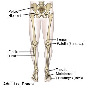
A leg is made up of three bones.
A leg is made up of three bones. One way to learn all the bones in the human body is to categorize them by shape. Diagram of the bones in the lower leg. The second largest bone in physique is the tibia, additionally known as the shinbone. Start learning with our skeleton diagrams, bone labeling exercises and skeletal system quizzes! What does this suggest about mammals? Also, defective and old red blood cells are destroyed in bone marrow. Dry docked u2013 three steps to avoiding ankle injuries. Most of the animals have the same bones, although some are shaped differently and placed in different positions. Cancellous bone produces red blood cells, platelets, and white blood cells. Tremendous advantages have been gained from this erect posture, the chief among which has been the freeing of the arms for a great. You can specify conditions of storing and accessing cookies in your browser. This quiz on human bones is designed to test your knowledge on the location of each individual bone.
You can specify conditions of storing and accessing cookies in your browser. They are very large and strong and help support the weight of your body. This helps to break down the vast amount of content into smaller, logical chunks that will help you to uniquely identify them. The foot bones shown in this diagram are the talus, navicular, cuneiform, cuboid, metatarsals and calcaneus. The calcaneus (heel bone) is the largest bone in the foot.
Leg Fracture Aftercare Instructions What You Need To Know from www.drugs.com Learn how to draw the femur, patella, tibia, and fibula in this lesson! Cancellous bone produces red blood cells, platelets, and white blood cells. Your legs are an amazing collection of bones and muscles. Foot bones images stock photos u0026 vectors. Diagram depicting the arterial supply to a growing leg. Since leg bones are important to our body structure, biomedical engineers design prosthetic legs to the strongest and largest joint in your body, the knee lets you move your lower leg back and forth as well good front and back human body skeleton diagram with bones identified. Bones in the foot powerpoint diagram. Tremendous advantages have been gained from this erect posture, the chief among which has been the freeing of the arms for a great.
They are connected to a group of bones called the pelvis , which supports the femur =the main bone in the upper part of your leg.
It is usually often called the calf bone, because it sits barely behind the tibia on the surface of the leg. But if something goes a bit wrong, they can hurt and make it hard to move around. Continue scrolling to read more below. Read or download foot bones diagram for free bones diagram at givediagram.marcopoli.it. However, the leg bone will now be parented in pose mode. You can specify conditions of storing and accessing cookies in your browser. Most of the animals have the same bones, although some are shaped differently and placed in different positions. Tremendous advantages have been gained from this erect posture, the chief among which has been the freeing of the arms for a great. Bone on hand and foot diagram quiz. Bones in the foot powerpoint diagram. Even when i parent the bones manually, both in offset and connected mode, the torso simply does not move the leg/hip bones. Learn how to draw the femur, patella, tibia, and fibula in this lesson! The bones of the leg are the femur, tibia, fibula and patella.
A diagram of the human skeleton showing bone and cartilage leg bones diagram. Foot bones images stock photos u0026 vectors.


0 Komentar